Every couple dreams of having a healthy baby. But sometimes, genetic conditions or fertility challenges stand in the way. However, advanced medical technology has developed methods for identifying high-risk cases.
This is where embryo biopsy helps.
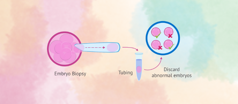
Embryo biopsy is a procedure used during IVF. It helps check for genetic disorders before implantation. Embryologists take a few cells from an embryo and test them. This can prevent the passing of inherited diseases.
There are different types of embryo biopsy techniques. Each has a unique approach. Some are done at an early stage, while others are later. Choosing the right type of embryo biopsy depends on the situation.
For many families, this procedure brings hope. Embryo biopsy increases the chances of a healthy pregnancy.
In this blog, we will explore the different embryo biopsy techniques, how they work, and why they are important.
- Understanding the Embryo Biopsy Procedure
- What Embryo Biopsy?
Embryo biopsy is a medical procedure used in in vitro fertilization (IVF) to check for genetic or chromosomal abnormalities. It involves removing a few cells from an embryo for genetic testing before implantation. This helps doctors select the healthiest embryo to increase the chances of a successful pregnancy.
- Step-by-step Explanation of How Embryo Biopsy is Performed
- Embryo Development
After fertilization, embryos grow in the lab. They develop for 3 to 5 days before the biopsy.
- Cell Removal
An Embryologist carefully extracts a few cells from an expanded embryo to a fully developed embryo without harming it.
- Genetic Testing
The extracted cells go through genetic analysis. Doctors check for chromosomal abnormalities or inherited diseases.
- Embryo Freezing
While waiting for results, embryos are frozen. This keeps them safe until transfer.
- Selecting the Best Embryo
Once testing is complete, doctors choose the healthiest embryo to proceed with the embryo transfer.
Embryo biopsy is a safe, effective, and precise procedure for increasing the chance of a successful pregnancy.
- Types of Embryo Biopsy Techniques
- Blastomere Biopsy Technique
- What is Blastomere Biopsy?
Blastomere biopsy is a preimplantation genetic testing technique where a single cell (blastomere) is extracted from a developing embryo at the cleavage stage. This procedure helps in diagnosing genetic disorders before implantation to improve the chances of a healthy pregnancy through in vitro fertilization (IVF).
- When it is Performed (Day 3 of embryo development)
This biopsy is performed on Day 3 of embryo development when the embryo has reached the 6—to 8-cell stage. At this stage, the blastomeres are loosely connected. This allows embryologists to safely extract a single cell without affecting the embryo’s viability.
- Advantages of Blastomere Biopsy Technique:
- Allows early genetic screening.
- Helps select healthy embryos before transfer.
- Increases the chance of a successful pregnancy.
- Challenges of Blastomere Biopsy Technique:
- Embryo viability concerns
- Some embryos may not develop to blastocyst or fully developed stage after a biopsy.
- Accuracy may be lower than later-stage biopsies.
- Trophectoderm Biopsy Technique
- What Is Trophectoderm Biopsy?
Trophectoderm (TE) biopsy is a technique in assisted reproductive technology (ART) where 5 to 10 cells are taken from the outer layer of a blastocyst (trophectoderm) for genetic testing (PGT).
- When It Is Performed (Day 5 Or 6 Of Embryo Development)
This biopsy is done on Day 5 or Day 6 of embryo development when the embryo has reached the blastocyst stage. At this stage, the embryo has over 100 cells. This makes it safer to remove a few without harming the embryo’s growth.
- Advantages Of Trophectoderm Biopsy Technique:
- Provides high accuracy in genetic testing.
- Reduces the risk of transferring embryos with genetic abnormalities.
- Improves the chances of a successful pregnancy.
- Does not harm the embryo’s inner cell mass, which develops into the baby.
- Challenges Of Trophectoderm Biopsy Technique:
- Requires advanced lab facilities and skilled specialists.
- Not all embryos develop into blastocysts.
- Slight risk of embryo damage, though it is minimal with experienced professionals.
- Polar Body Biopsy Technique
- What Is a Polar Body Biopsy?
Polar body biopsy is a genetic testing method performed on a woman’s egg before fertilization. It examines the small cells (polar bodies) an egg (oocyte) releases during its development. Since polar bodies contain genetic material from the egg, they can indicate whether the egg has normal or abnormal chromosomes.
- When it is Performed (Before Embryo Formation, During Egg Development).
This technique is performed during egg development, before fertilization. It can be done in two stages:
- Simultaneous Biopsy: Both the first and second polar bodies are removed 6-14 hours after fertilization.
- Sequential Biopsy: The first PB is taken before fertilization, and the second PB is taken after fertilization.
- Advantages of the Polar Body Biopsy Technique
- Non-invasive to the embryo
- Early genetic screening
- Reduces risk of genetic disorders
- Challenges of the Polar Body Biopsy Technique
- Limited genetic insight
- Not suitable for all genetic conditions
- Lower accuracy compared to embryo biopsy
- Embryo Genetic Testing Methods Used After Biopsy
- Overview of Preimplantation Genetic Testing (PGT)
Preimplantation Genetic Testing (PGT) is a procedure used to analyze the genetic makeup of an embryo before implantation. After a biopsy, the extracted cells are tested to detect potential genetic issues. This helps in selecting healthy embryos for transfer during IVF to reduce the risk of genetic disorders and improve healthy pregnancy success rates.
- Types of genetic testing done on biopsied cells:
- PGT-A (Aneuploidy Screening)
This test checks for missing or extra segments of chromosomes or complete chromosomes. Chromosomal abnormalities can lead to failed implantation, miscarriage, or genetic disorders like Down syndrome.
- PGT-M (Monogenic Disorder Testing)
It identifies specific inherited diseases caused by single gene mutations. This is helpful for couples with a family history of genetic conditions like cystic fibrosis or sickle cell anemia.
- PGT-SR (Structural Rearrangements)
This test detects chromosome structural changes, such as missing or rearranged parts. These issues can cause infertility, miscarriages, or congenital disabilities.
Secure a Healthy Pregnancy with the Right Testing
Embryo genetic testing supports a healthier pregnancy by selecting embryos with the best chances of success. Different biopsy techniques help collect essential genetic information while keeping the embryo safe. Doctors carefully choose the best approach for each patient because precision, safety, and successful outcomes matter to us.
With advancements in embryo biopsy techniques, fertility treatments are becoming more effective.
If you are considering genetic testing, talk to a fertility specialist at Motherhood Hospital.
As the best embryo biopsy center in Ahmedabad, we provide advanced techniques to ensure accurate results.
Our passionate team ensures precision care, safety, and state-of-the-art technology, a patient-focused approach with the highest chances of success for every hopeful parent. Trust Motherhood Hospital to provide expert fertility care because your hopes, future, and little one’s first step matter to us.